Cornell Medical Instruments - Fundus Camera and Retinal Imaging Devices
Monaco

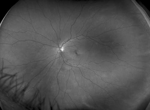
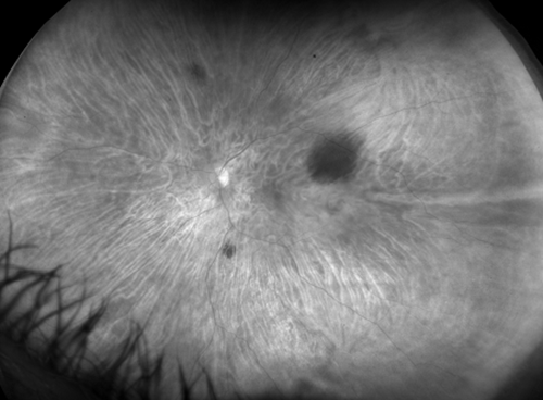
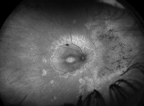
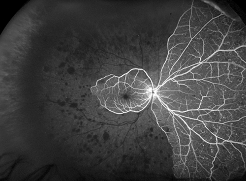
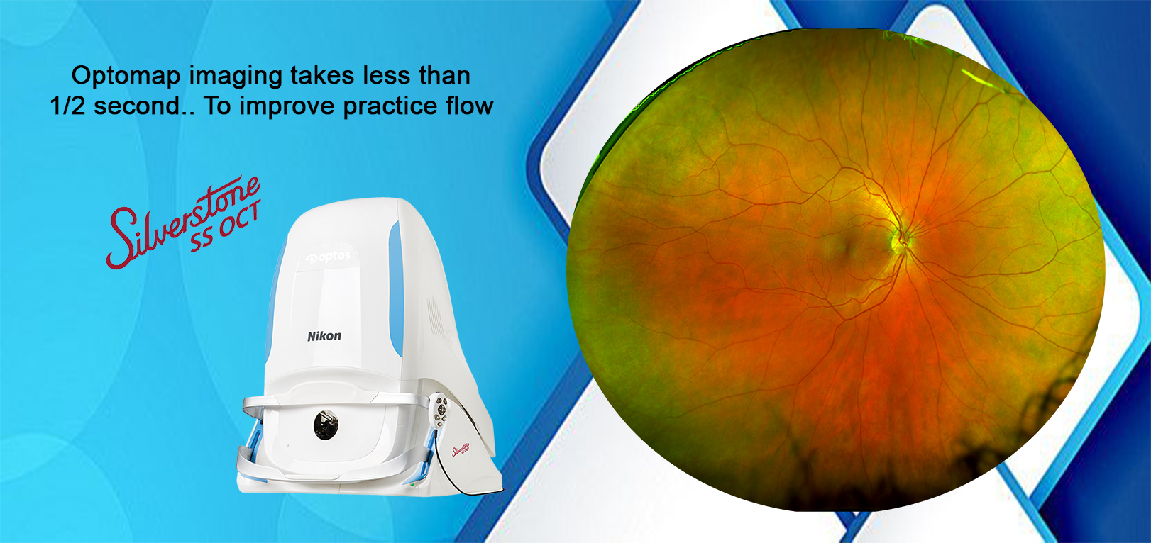
Monaco The 200 Degree, Single Capture Retinal Imaging System With Integrated OCT, Which Shows Virtually The Entire Retina In Less Than ½ Second And Provides Cross-sectional OCT Views. 

Optomap color

Red Free

Choroidal

Optomap af

OCT

California

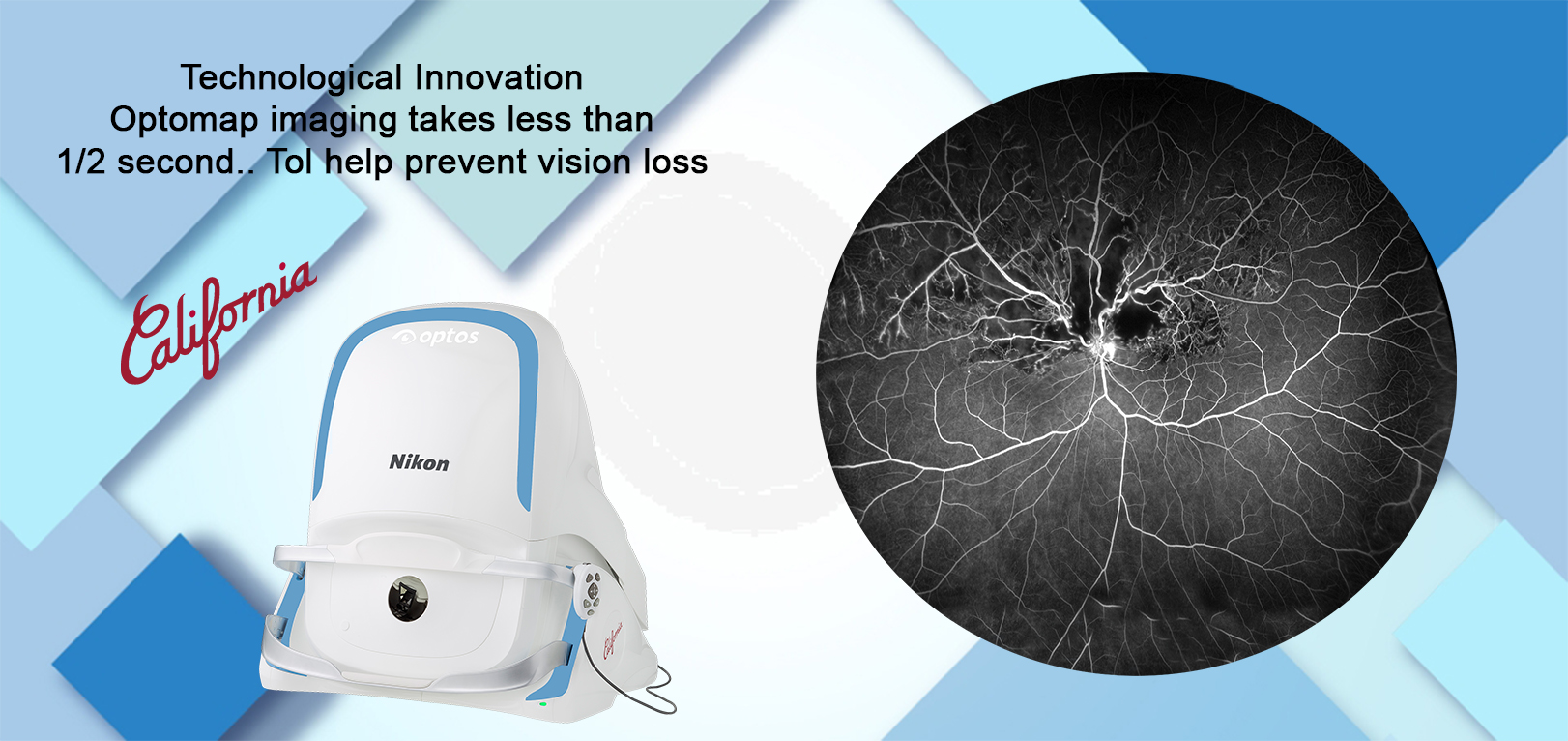
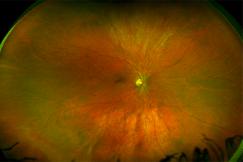
California Has Three Versions Available To Be Customizable For Use In Multiple Eyecare Settings And The New Hardware And Software Technology Will Enable Practitioners To See More, Discover More And Effectively Treat More Ocular Pathology Thus Promoting Patient Health.

Color

Red-Free

Choroidal

Autofluorescence
California Fa

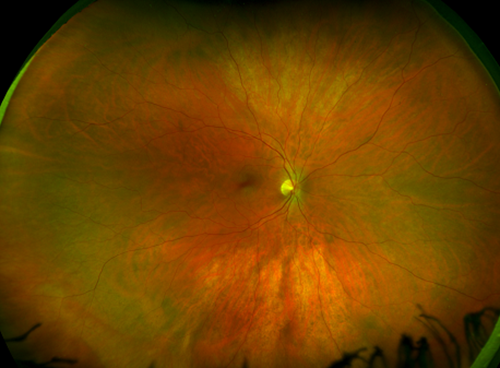
California Has Three Versions Available To Be Customizable For Use In Multiple Eyecare Settings And The New Hardware And Software Technology Will Enable Practitioners To See More, Discover More And Effectively Treat More Ocular Pathology Thus Promoting Patient Health.

Color

Red Free

Choroidal

Autofluorescence

Fluorescein Angiography
California Icg

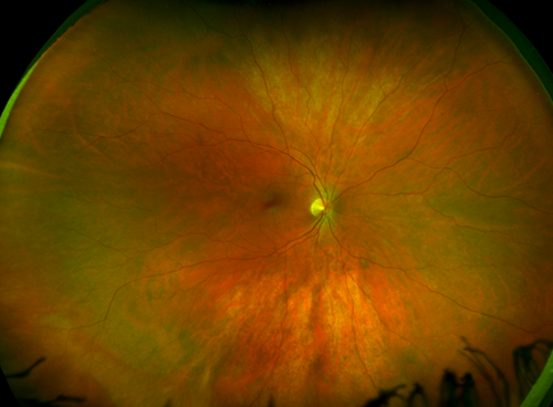
California Has Three Versions Available To Be Customizable For Use In Multiple Eyecare Settings And The New Hardware And Software Technology Will Enable Practitioners To See More, Discover More And Effectively Treat More Ocular Pathology Thus Promoting Patient Health.

Color

Red Free

Choroidal

Autofluorescence
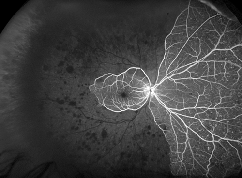
Fluorescein Angiography
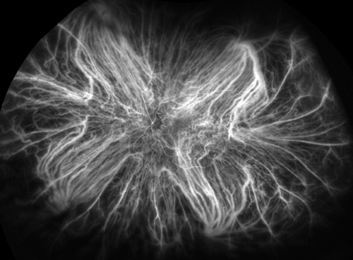
Indocyanine Green Angiography

Daytona NG

Daytona NG Provides Eyecare Professionals With UWF Digital Images Of 200 Degrees Or Up To 82% Of The Retina In A Single, Non-contact Optomap Image. In Addition, The Daytona NG Device Comes With The OptosAdvance™ Browser-based Image Review Software, Which Allows For Simple Documentation, Monitoring, And Referral Processing To Assist In Patient Management And Improved Patient Flow.
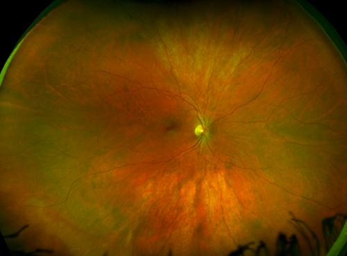
Color
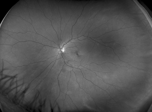
Red Free
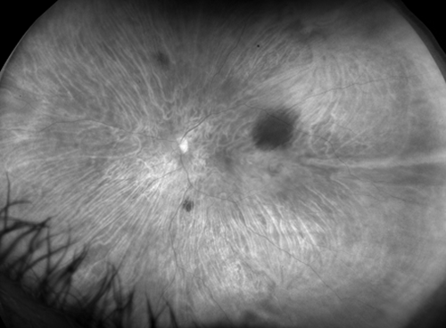
Choroidal
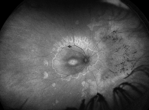
Autofluorescence
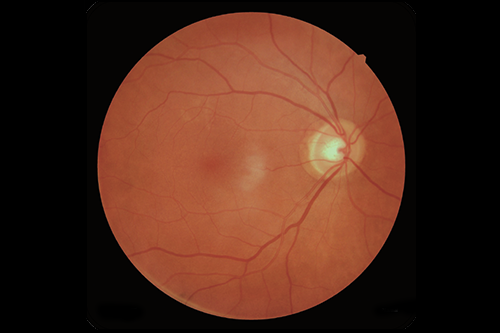
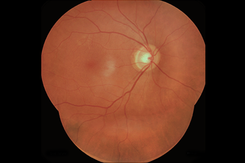
RetinaStation

The RetinaStation System Is A Fully-automatic, Non-mydriatic 45° Retinal Imaging Devicewith An Eye To The 3 Smart Features.For Practitioners, The New Model Is Well Thought-out In Many Ways.

ECLIPSE Ti2 Series

Nikon Inverted Microscopes Are Renowned For Optical Quality, Flexibility, Modularity, Ease Of Use, And Mechanical Precision. Serving As Either As A Standalone System Or By Powering The Core Of Complex, Multimodal Imaging Systems, Nikon’s Inverted Microscopes Ensure The Highest Imaging Results For Every Experiment.  

Ti2-A

Ti2-E

Ti2-U

ECLIPSE Ts2R

Compact Inverted Research Microscope Configurable With A Wide Variety Of Observation Methods

ECLIPSE Ts2R

TS2R-Stage

ECLIPSE Ts2

Nikon's Inverted Routine Microscope Featuring A Compact Footprint, Newly Developed Contrast Observation Method And Improved Operability, Ideal For The Tissue Culture Lab

ECLIPSE Ti2-U IVF

This Product Is Available For Clinical Use In The United States. Please Contact Your Local Nikon Dealer For Information On Clinical Use Of This Product In Your Country.

ECLIPSE Ni Series

Research And Clinical Upright Microscopes That Support Research In The Fields Of Biological Science And Medicine, With Excellent Optical Performance And High System Expandability That Can Be Equipped With Motorized Accessories.

NI-E_Trinoc

NI-L

NI-E

ECLIPSE Ci Series

Ergonomic Upright Microscopes With Eco-illumination For Clinical And Laboratory Applications.

CI-E

Ci-E1

Ci-L

Ci-S

ECLIPSE FN1

Upright Microscope With A Fixed Stage And Focusing Nosepiece Dedicated To Electrophysiological Research.

Eclipse Fn1

Sensapex FN-M

Electrophysiological Research Microscope Featuring Single Or Dual Motorized Nosepieces For Smooth Objective Switching While Maintaining Sample Positioning And Focus.


Sensapex FN-M

Sensapex_FNM

ECLIPSE Si

Ergonomically Designed Nikon's Upright Microscope, ECLIPSE Si Brings Comfort And Precision To Your Operation With Cutting-edge Hardware And Software Solutions.

ECLIPSE-Si

ECLIPSE Ei

The ECLIPSE Ei Educational Microscope Offers Unique Digital And Design Solutions To Ensure The Smooth Progress Of Science And Technology.

ECLIPSE Ei

ECLIPSE LV100N POL LED

The Highest Level Of Optical Quality, Operability And Stability For Polarized Light Microscopy. It Is Equipped With A Bright LED Light Source For Minimized Heat-induced Focus Drift. This Product Is Suitable For A Wide Range Of Imaging Applications.

ECILIPSE LV

ECLIPSE Ci POL

Compact Polarizing Microscope Featuring Superb Optical Performance And Ease-of-use.

ECLIPSE Ci POL

SMZ25 / SMZ18

Breakthrough Stereo Microscope Optics Delivering High Zoom Ratio Of 25:1, Unprecedented Resolution And Bright, Fluorescent Images. Capture Stunning Images And Time-lapse Data With Incredible Detail And Field Of View.

SMZ2518

SMZ1270 / SMZ1270i

Stereo Microscopes Offering An Impressive 12.7:1 Zoom Ratio, High Resolution Optics, And Enhanced Ergonomics.

SMZ1270 / SMZ1270i

SMZ800N

Affordable Model With 8:1 Zoom Ratio, Streamlined Operation And Robust Performance.

SMZ800N

SMZ745 / SMZ745T

Stereo Microscope With 7.5:1 Zoom Ratio With Trinocular Head Option For Digital Imaging. Features One Of The Highest Zoom Magnifications In Its Class.

SMZ745 / SMZ745T

SMZ445 / SMZ460

Compact, Lightweight And Cost Effective Stereo Microscopes Featuring Porro Prism Optics.

SMZ445 / SMZ460

Stereo Microscope Accessories

A Variety Of Accessories For Stereoscopic Observations, Including Objectives, Observation Tubes, Illuminators, Stands And Stages.

Stereo Microscope Accessories

Digital Sight 50M

60.0-megapixel, High-definition, Cooled Monochrome Camera That Combines Large Field Of View And Fast Frame Rates.

Digital Sight 50M

Digital Sight 10

High Resolution Of 6K And Switching Color And Monochrome Capture With A Single Camera, Also Features A High Frame Rate For Fast Focusing On High-definition Images.

Digital Sight 10

Digital Sight 1000

2.0-megapixel Color Camera Offering Full-HD Images, Fast Live Display And Simple Measurement, Without A PC.

Digital Sight 1000

DS-Fi3

5.9-megapixel Color Camera Offering Low-noise, High-resolution Imaging, Fast Live Display And Exceptional Color Reproduction.

DS-Fi3

NIS-Elements L

Tablet-based Controller For Digital Sight 10, Digital Sight 1000 And DS-Fi3. Streamlined Control And Simple Measurement And Annotation Tools.

NIS-Elements L

X-Light V3

Spinning Disk Confocal Revolutionising The Visualization Of Biologic Sample All At Once


X-Light V3

X-Light V2

UNIVERSAL PLUG-IN SPINNING DISK FOR FAST AND GENTLE IMAGING

X-Light V2

CICERO

YOUR CONFOCAL JOURNEY STARTS HERE
A Flexible, Compact But Full-fledged Spinning-disk Confocal Solution, CICERO Makes High-end Fluorescence Imaging Accessible To Every Laboratory Enabling A Variety Of Applications


CICERO

Silverstone

Silverstone produces A 200° Single-capture Retinal Image Of Unrivaled Clarity In Less Than ½ Second And Enables map- OCT Scanning Across The Retina And Into The Far Periphery. 

optomap sensory-red-free

Optomap-choroidal

-optomap-color-rg

Optomap-fa

Optomap-green-af-1

Optomap-icg

Swept-source-oct

SilverStone Device

PANTHEON

AI-driven Ecosystem Transforming Pathology

QAi PROSTATE

An Intuitive AI-powered Ecosystem Which Digitizes The Entire Workflow For Pathologists From Start To Finish – Making Way For The Possibilities Of Remote Diagnosis And Telepathology. It Enhances Concordance And Patient Diagnosis, Allowing Pathologists To Focus More Time On Complex Cases.

QCMOS

The World's First Camera To Incorporate The QCMOS Image Sensor. The Camera Achieves The Ultimate In Quantitative Imaging.

1(a)

1(b)

2

3.(a)

3.(b)

4.(a)

4.(b)

SCMOS

A Leader In The Scientific CMOS Camera Revolution, Hamamatsu Has A Camera To Fit Almost Any Budget Or Application.

InGaAs

InGaAs Cameras Bridge The Gap Between NIR Wavelengths In The 950-1700 Nm Range, Where Silicon Detectors Are No Longer Sensitive. Our Products Capture Images With QVGA To VGA Resolution And Our Extensive Experience With InGaAs Sensors Allows Us To Offer Cameras With Exquisite Image Contrast And Quality.

TDI

High-speed Imaging Of Moving Samples Is A Challenging Even In Bright Conditions, But Our TDI Cameras Turn Linear Movement Of The Sample Into An Advantage By Coordinating Signal Accumulation In The Sensor With Sample Movement. These Cameras Are Also Well-suited For Low-light Scanning Applications That Are Too Dim To For A Line Scan Sensor.

PE-800

8-channel Illumination And Lightning Fast Control

Spinal Cord labelled with DAPI/EGFP-Lamnin-GFAP (pE-800)

Enteric Glia Cells (Green) and Endothelial Cells (Red), acquired using the CoolLED (pE-800)

Enteric Nervous System labelled with GFAP and HuCD (pE-800)

Primary neuron from a rat cortex – supplied by Transnetyx Tissue by BrainBits (CoolLED pE-800)

Bovine pulmonary epithelial cells. (CoolLED pE-800)

PE-800fura

Building On The Successful PE-800 Illumination System, The PE-800fura Offers The Most Comprehensive Calcium Imaging System Available. It Also Enhances A Range Of Applications From PH Monitoring And Optogenetics To Everyday Fluorescence Microscopy, Combining Lightning-fast Switching With Eight Individually Controllable Channels. Backed By CoolLED’s World-renowned Support And Generous Warranty, The PE-800fura Presents The Go-to Illuminator For Calcium Imaging And Beyond.

CoolLED pE-800fura

PE-400

Building On The Success Of The Popular PE-300 Series, The PE-400 Is A Simple Bright White Light Illumination System And A Controllable, Cost-effective Replacement For Mercury And Metal Halide Lamps. Four Powerful LEDs Offer Intense, Broad-spectrum Coverage For The Most Popular Fluorophores, Ranging From DAPI Through YFP To Cy5.

PE-400max

Building On The Success Of The Popular PE-300 Series, The PE-400max is The Most Advanced Illumination System Of The PE-400 Series. Four Powerful LEDs Can Be Individually Controlled Via Software, TTL, Lightbridge GUI And Manual Control Pod For Demanding Everyday Fluorescence Microscopy And Optogenetics Applications Using The Most Popular Fluorophores, Ranging From DAPI Through YFP To Cy5.

Mouse brain section stained with anti-GFP (pE-400max)

PE-4000

The PE-4000 Sets The Standard As The Universal Illumination System For Fluorescence Microscopy. The System Has 16 Selectable LED Wavelengths Across Four Channels That Can Be Finely Controlled And Matched To The Filters And Fluorophores Of Almost Any Microscope, Making It The Broadest And Most Versatile Illumination System Available.

The CoolLED PE-4000 Benefits From Our Award Winning Sustainable Green Technology And Delivers Enhanced Irradiance At The Sample Plane With A Significant Reduction In Energy Consumption, And Is Supplied With A 36 Month Warranty.

Zebrafish larvae within the field of the Mesolens. (CoolLED pE-4000)

A GFP-labelled keratinocyte undergoing mitosis. (CoolLED pE-4000)

Time series of keratinocytes migrating during live-cell imaging outgrowth assay. (CoolLED pE-4000)

Cultured hippocampal neurons. (CoolLED pE-4000)

PE-300lite

CoolLED’s PE-300lite is A Simple White Light LED Illumination System That Offers Intense, Broad-spectrum LED Illumination For Fluorescence Microscopy. The System Is Perfect For Hospital And Regular Research Laboratory Environments And Is Affordable Within Consumables Budgets.
This Mercury-free Alternative Is Safer, More Controllable And Precise Than Conventional Bulb Illumination Options. Spectral Coverage Is From The UV (DAPI Excitation) To The Red Region (Cy5 Excitation).

LED Technology Is Quickly Evolving, And Since 2017 Our New Generation Has Double The Irradiance At The Sample Plane, And For Even Higher Irradiance Is Available As A direct Fit Model (which Also Reduces The Cost To Buy And Run Thanks To Fewer Consumables). With ACT Label Certification, It’s Even An Environmentally Friendly Option For Illumination.

PE-300white

CoolLED’s PE-300white Offers Intense, Broad-spectrum LED Illumination For Fluorescence Microscopy That Is Perfect For Hospital And Regular Research Laboratory Environments. The System Is A Mercury-free Alternative That Is Safer, More Controllable And Precise Than Traditional Bulb Illumination Options.  Spectral Coverage Is From The UV (DAPI Excitation) To The Red Region (Cy5 Excitation).
 
LED Technology Is Quickly Evolving, And Since 2017 Our New Generation Has Double The Irradiance At The Sample Plane, And For Even Higher Irradiance Is Available As A Direct Fit Model (which Also Reduces The Cost To Buy And Run Thanks To Fewer Consumables). With ACT Label Certification, It’s Even An Environmentally Friendly Option For Illumination.

E18 Sprague Dawley Hippocampus grown out to DIC 8 – supplied by Transnetyx Tissue by BrainBits (CoolLED pE-300white)

E18 Sprague Dawley Hippocampus grown out to DIC 8 – supplied by Transnetyx Tissue by BrainBits (CoolLED pE-300white)

E18 Sprague Dawley Hippocampus grown out to DIC 8 – supplied by Transnetyx Tissue by BrainBits (CoolLED pE-300white)

Multi-stained skin cells. (CoolLED pE-300white)

PE-300ultra

The CoolLED PE-300ultra Is A Highly Controllable Fluorescence Microscopy Illumination System Which Offers Intense, Broad-spectrum LED Illumination For Imaging Most Common Fluorophores.  Fast Events Can Be Captured With Microsecond Switching Via Multiple TTL Inputs, And The Ability To Mount Inline Excitation Filters Allows Fast, Flexible Filter Configurations.  This, When Paired With Today’s High-performance Multi-band Filter Sets, Facilitates Imaging Traditionally Done Via A White Light Source And A Filter Wheel, With All The Benefits Of LEDs.
 
LED Technology Is Quickly Evolving, And Since 2017 Our New Generation Has Double The Irradiance At The Sample Plane, And For Even Higher Irradiance Is Available As A Direct Fit Model (which Also Reduces The Cost To Buy And Run Thanks To Fewer Consumables). With ACT Label Certification, It’s Even An Environmentally Friendly Option For Illumination.

Malaghan Institute – Cas9Ccl17-eGFP expressing bone marrow cells (pE-300ultra)

Triple-stained fixed HeLa cells overlaid with holotomography. (Nanolive/CoolLED pE-300ultra)

Live FUCCI mouse embryonic stem cells. (Nanolive/CoolLED pE-300ultra)

Live HeLa cells stained with lysosome marker. (Nanolive/CoolLED pE-300ultra)

PE-100

The PE-100 Series Is A Family Of Single-wavelength Fluorescence LED Illumination Systems, Which Are Compact And Simple To Use.  Systems Can Be Specified At Any One Of 20 Different LED Wavelengths, And Are Ideal For Routine Clinical Screening, Electrophysiology Or Basic Research Applications. Operation Is By A Remote Manual Control Pod With Instant On/off And Irradiance Control From 0-100%, With Remote Control Available Via A TTL Trigger.

Huwe1 protein localisation in human bronchial epithelial cells.

Neurospora crassa hyphal filaments.

Aspergillus fumigatus germlings.

Aspergillus fumigatus microcolony on an epithelial alveolar monolayer.

PT-100

The PT-100 Is A Compact And Simple-to-use LED Illumination System For Transmitted Light Applications, Including Brightfield, Darkfield, DIC, Dodt Gradient Contrast And Phase Contrast.
 
The System Is Available In Four Variants To Bring The Benefits Of LEDs To Modern Laboratories. A Broad White System Provides A Safer And Convenient Alternative To Halogen By Reducing Harmful UV Exposure To The Eyes And Is Ideal For Long Hours Spent At The Microscope, While Three Narrow Bandwidth Options (525, 635 And 770 Nm) Optimise Image Quality Through Increased Contrast. The PT-100-WHT And PT-100-770 Nm Are Also Available As Variants With A 3 Mm Liquid Light Guide Attachment, Which Is Ideal For Electrophysiology Applications.


PT-100-LLG-1800-x-1800-px-300x300

PT-100-Pod-1800-x-1800-px-150x150

Transmitted Podcarpus Elatus Leaf image. (CoolLED pT-100-WHT)

Living human oocytes. (CoolLED pT-100)

TissueFAXS Q & SL Q

TissueFAXS Q & SL Q Systems
Confocal Tissue Cytometer

TissueFAXS Q & SL Q Systems Provide The Benefits Of Both High-throughput Of Slides And Laser-free Fast Confocal Imaging. All TissueFAXS Fluo Or PLUS System Configurations, Irrespective Of Whether They Are Upright Or Inverted, Are Also Available As TissueFAXS Q Configurations.

TissueFAXS SPECTRA

TissueFAXS SPECTRA 
Multispectral Tissue Cytometer

TissueFAXS SPECTRA Systems Provide Multispectral Imaging, Spectral Unmixing And Contextual Tissue Cytometry Image Analysis On The Basis Of The TissueFAXS Platform.

SPECTRA Technology Is Available On Any TissueFAXS System Configuration, Be It Upright, Inverted, TissueFAXS Histo, TissueFAXS Fluo, TissueFAXS PLUS Or Slide Loader-based (SL).

TissueFAXS CHROMA

TissueFAXS CHROMA 
High-Speed Multispectral Tissue Cytometer

TissueFAXS CHROMA Specializes In Automated Multispectral Fluorescence Whole Slide Scanning For Up To 7 Markers At A Time Made Possible By An Optimized Filter Set Eliminating Channel Bleed Through. This Cost-effective High-speed Slide Scanner Reaches Its Full Potential When Combined With Single Cell And Contextual Image Analysis. This Platform Is A Holistic Solution For Cellular Phenotyping And In-depth Tissue Microenvironment Characterization Of IF Processed Tissue Sections, TMAs, And Cells On Standard And Oversized Slides.

TissueFAXS SL

TissueFAXS SL
SLIDE LOADER

All TissueFAXS Upright Systems Configurations (Fluo, Histo, PLUS) Are Also Available As TissueFAXS SL (Slide Loader) High-throughput Systems.

In This Configuration, They Are Additionally Equipped With An Automatic Slide Loader And A Specially Adapted Version Of The TissueFAXS Scanning And Image Management Software.

TissueFAXS PLUS

TissueFAXS PLUS 
UPRIGHT BRIGHTFIELD AND FLUORESCENCE SYSTEM

TissueFAXS PLUS Is An Upright Fluorescence And Brightfileld System For The Scanning And Analysis Of Slides, Cytospins, Smears And Tissue Microarrays. The System Is Equipped With TissueQuest/HistoQuest Or StrataQuest PLUS Image Analysis Software.

TissueFAXS I PLUS

TissueFAXS I PLUS 
INVERTED BRIGHTFIELD AND FLUORESCENCE SYSTEM

TissueFAXS I PLUS Is An Inverted Fluorescence And Brightfield System For The Scanning And Analysis Of Slides, Cytospins, Smears And Tissue Microarrays. The System Is Equipped With TissueQuest/HistoQuest Or StrataQuest PLUS Image Analysis Software.

TissueFAXS Fluo

TissueFAXS Fluo
UPRIGHT FLUORESCENCE SYSTEM

TissueFAXS Fluo Is An Upright Fluorescence-only System For The Scanning And Analysis Of Slides, Cytospins, Smears And Tissue Microarrays. The System Is Equipped With Either TissueQuest Or StrataQuest Fluo Image Analysis Software

TissueFAXS I FLUO

TissueFAXS I Fluo 
INVERTED FLUORESCENCE SYSTEM

TissueFAXS I Fluo Is An Inverted Fluorescence System For The Scanning And Analysis Of Slides, Cytospins, Smears And Tissue Microarrays. The System Is Equipped With TissueQuest And/or StrataQuest Fluo Image Analysis Software.

TissueFAXS Histo

TissueFAXS Histo 
UPRIGHT BRIGHTFIELD SYSTEM

TissueFAXS Histo Is An Upright Brightfield Only System For The Scanning And Analysis Of Slides, Cytospins, Smears And Tissue Microarrays. The System Is Equipped With HistoQuest And/or StrataQuest Histo Image Analysis Solftware.

TissueFAXS I HISTO

TissueFAXS I Histo 
INVERTED BRIGHTFIELD SYSTEM

TissueFAXS I Histo is An Inverted Brightfield System For The Scanning And Analysis Of Slides, Cytospins, Smears And Tissue Microarrays. The System Is Equipped With HistoQuest or StrataQuest Histo image Analysis Software.

StrataFAXS II

StrataFAXS II 
Streamlined Tissue Cytometry

StrataFAXS II is An Upright Brightfield Slide Scanner For acquisition and Analysis Of HE/IHC Processed Tissue Sections, Smears and Tissue Microarrays (TMA). StrataFAXS II With Its Innovative Workflow Allows You To Directly Work On Your Samples Via Real-time Scanning And Live Stitching. The System Is Equipped With HistoQuest Single Cell Analysis Software And/or StrataQuest Contextual Image Cytometry Software Which Includes A Machine Learning Module For Automated Classification Of Histological Entities (e.g. Tumor Foci, Epidermis, Glands, Glomeruli) And A Deep Neural Network For Precise Nuclear Segmentation Even At High Cellular Densities And Low Expression Levels.

THMS600

Temperature And Environmental Control From < -195°C To 600°C With XY Sample Manipulators

THMSG600

Temperature And Environmental Control From < -195°C To 600°C Optimised For Earth Sciences

THMSEL600

THMS600 Ellipsometry Stage, Optimised For Use With Ellipsometers

THMS600-PS

Temperature And Environmental Control From -100°C To 500°C At High Pressure

FTIR600

Temperature And Environmental Control From < -195°C To 600°C, Optimised For Infrared And FTIR Systems

Optical DSC600

Single Cell Calorimetry Optimised For Imaging And Spectroscopy, Temperature Control From < -195°C To 600°C

HFS600

Temperature And Environmental Control From < -195°C To 600°C

HFS600E-P

Temperature And Environmental Control From < -195°C To 600°C With Options For Probes And Electrical Connections

MFS - Modular Force Stage

Mechanical Sample Characterisation With Temperature And Environmental Control Modules

MDS600 Motorised System

Temperature And Environmental Control From < -195°C To 600°C With Motorised XY Sample Manipulators

MDSG600 - Motorised Geology System

Temperature And Environmental Control From < -195°C To 600°C With Motorised XY Sample Manipulators, Optimised For Earth Sciences

Cap500

Temperature Controlled Capillary Stage From < -195°C Up To 500°C

LTS420

Temperature And Environmental Control From < -195°C To 420°C

LTS420E-P

Temperature And Environmental Control From < -195°C To 420°C With Options For Probes And Electrical Connections

THMS350V

  • Temperature Range < -195°C* To 350°C (*with LNP96)

THMSEL350V

Temperature Control Vacuum Stage For Ellipsometry Systems

HFSX350

Systems For X-ray, XRD And Beamline Applications

HFS350EV-PB4

Temperature And Vacuum Control From < -195°C To 350°C With Options For Probes And Electrical Connections

CSS450

Optical Rheology System

DSC450

Differential Scanning Calorimetry With Imaging, Temperature Control From < -150°C To 450°C

FDCS196 Freeze Drying System

BCS196 Cryobiology System

MDBCS196

Temperature And Environmental Control From < -195°C To 125°C With Motorised XY Sample Manipulators

LTS120

Temperature And Environmental Control From -40°C* To 120°C

LTS120E-P ELECTRICAL & PROBE SYSTEM

TS1000

Temperature And Environmental Control From Ambient To 1000°C

TSEL1000

TSEL1000 Ellipsometry Stage, Optimised For Use With Ellipsometers

TS1000E-PB4

Temperature And Environmental Control From Ambient To 1000°C With Probes And Electrical Connections

CCR1000 - Catalysis Cell Reactor

Temperature And Environmental Control From Ambient To 1000°C

TS1200

Temperature And Environmental Control From Ambient To 1200°C

TS1400XY

Temperature And Environmental Control From Ambient To 1400°C With XY Sample Manipulators

TS1500

Temperature And Environmental Control From Ambient To 1500°C

CMS196V4

Cryo-Correlative Microscopy Stage

Geological

Understanding And Characterising The Thermal Properties Of Your Sample Is Essential In A Variety Of Applications.

Semiconductors And Electrical

Understanding And Characterising The Thermal Properties Of Your Sample Is Essential In A Variety Of Applications.

Materials And Metallurgy

Understanding And Characterising The Thermal Properties Of Your Sample Is Essential In A Variety Of Applications.

Pharmaceutical

Understanding And Characterising The Thermal Properties Of Your Sample Is Essential In A Variety Of Applications.

Plastics And Polymer

Understanding And Characterising The Thermal Properties Of Your Sample Is Essential In A Variety Of Applications.

Oil & Gas

Understanding And Characterising The Thermal Properties Of Your Sample Is Essential In A Variety Of Applications.

Life Science

Understanding And Characterising The Thermal Properties Of Your Sample Is Essential In A Variety Of Applications.

Food Science

Understanding And Characterising The Thermal Properties Of Your Sample Is Essential In A Variety Of Applications.

Space Science

Understanding And Characterising The Thermal Properties Of Your Sample Is Essential In A Variety Of Applications.

What Our Clients Say
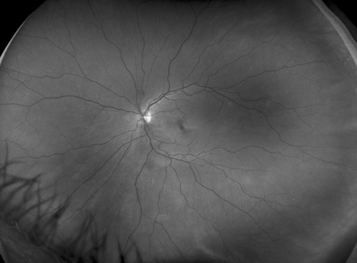
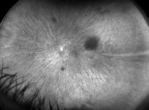
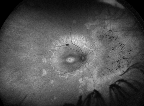
California Is Capable Of Performing UWF Fundus Photo, UWF-FAF, UWF-FFA And UWF-ICG. The Machine Is Very Easy To Setup And Use With An Impressive Interface Allowing Various Post Exam Modification To The Image. The Machine Provides Very Good Quality Ultra Wide Field Images Even In Undiliated Pupil.
We Could Capture Almost All The Peripheral Retinal Lesions Like Lattice Degeneration, Retinal Holes, Peripheral Neovascularisation, Peripheral Non Perfusion Areas, Peropheral Tumors Etc.

- Dr. Vikram Vinayak Koundanya

Daytona Is A Wonderful Machine To See Largest Field Of View In Single Shot, Non Mydriatic Imaging In Just 30 Seconds. We Have Seen Various Abnormalities At Peripheral Region Which We Were Unable To See With Regular Fundus Camera.

I Recommed That Optos Machine Has Significant Use For Patient Examination And Documentation.

- Dr. Amit Khosla

I Am Using Optos Daytona Since Last So Many Years. This Is The Best Equipment For Retinal Imaging And Capture Almost 82 Percent Of Retina In Just 30 Seconds. I No Longer Need To Dialate My Patients And Ask Them To Wait Before Taking Fundus Images. The Machine Software Keeps The History Of Patient To Compare It In Further Visits To Monitor The Progress. 

- Dr. Manoj Saswade

We Evaluated Optos California ICG Which Has The Capability Of Doing Wide-field Color, Red-free, Autofluorescence, Fluorscien Angiography And ICG Angiography. We Were Particularly Impressed With The Image Quality Obtained In Pediatric Patients With Uviites And Those Patients With ROP Who Had Received Treatment In Their Infancy. 

The Ease Of Doing Wide-field FFA In Children Was Very Impressive. We Were Able To Image Extreme Pheriphery Even In The ROP Children.

I Would Strongly Recommend Optos California To All Retinal, Uveitis Specialists And Pediatric Ophthalmologists.

- Dr. Alay Banker

Ask CMI for quotation