


Product Description
TissueFAXS SL
TECHNOLOGY
TissueFAXS SL integrates TissueGnostics Slide Validator technology to ensure a high level of digital slide sharpness. Each scanned FOV is evaluated for sharpness on-the-fly during the scan process.
If the validation algorithm deems the FOV to be not sharp enough it will be rescanned on-the-fly.
TissueFAXS SL
Workflow
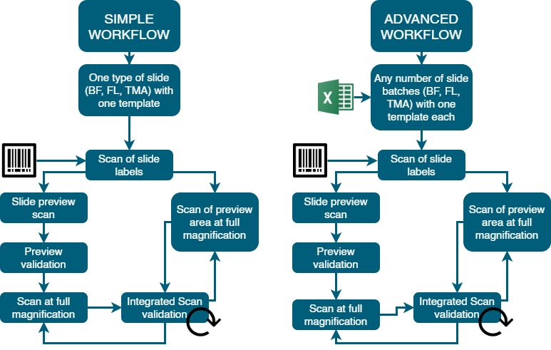
TissueFAXS SL comes with a new version of the TissueFAXS software designed for the perfect handling of the high amount of samples, data validation and storage. There are two main workflows, a simple workflow for one type of slides and an advanced workflow for mixed samples types:
Technical Specifications
